
Summary:
You have the scan, you wait, and then the report lands in your patient portal before anyone has had a chance to talk you through it. Within a few lines you are reading about desiccation, protrusion, foraminal narrowing and facet arthropathy. It reads like a list of things that are wrong with you.
Here is the part nobody explains up front. A radiologist is doing exactly the right job by describing every structural detail visible on the images. What they are not doing is deciding whether those details are the reason you hurt. That is a separate question, and it takes a clinician who has examined you, listened to your history and tested your reflexes to answer it.
Understanding that difference changes the entire conversation you are about to have.
What An MRI Actually Shows, And What It Cannot Show
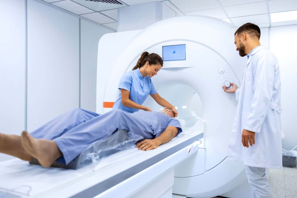
An MRI uses a strong magnetic field and radio waves to build cross sectional images of your spine. Unlike an X-ray, it shows soft tissue clearly, which means discs, nerve roots, the spinal cord, ligaments and the fluid surrounding them. That is why it is the imaging study of choice when a physician suspects nerve compression, a disc problem, or something less common that needs to be ruled out before treatment moves forward.
What an MRI does not show is pain. There is no sequence, no view and no setting that makes pain visible. It shows anatomy at one moment in time, in a person lying still on their back, which is very often not the position that hurts. Two people can have nearly identical images and completely different levels of function, comfort and capability. That gap between the picture and the lived experience is the single most useful thing to understand about your report.

Why Does My MRI Report Sound So Alarming?
Radiology reports are written physician to physician. The language is deliberately precise and deliberately neutral, but precision reads as severity when you are the person being described. A phrase such as moderate neural foraminal narrowing is a measurement, not a verdict. Words like degeneration and deterioration describe tissue that has changed over time, which is simply what tissue does. In everyday English those words sound like decline and damage. In radiology they sit much closer to describing gray hair or reading glasses.
The report also lists everything, by design. A radiologist who omits a finding, even a trivial one, has failed at the job. So they document every level of your spine, every disc, every small change, whether or not any of it has the slightest connection to why you came in. The result is a document that reads like a catalogue of problems when a great deal of it is ordinary background. You are looking at a complete inventory, not a summary of what matters.
There is also no narrative anywhere in the report. It does not say that the finding at L4 to L5 lines up neatly with the numbness you described running down the outside of your calf. It cannot say that, because the radiologist usually has only a one line clinical history and has never met you. Connecting the images to your story is the job of the physician who examined you, and that step is what turns a scan into a plan you can act on.
So if you read your report and felt your stomach drop, that reaction is completely normal, and it is not evidence that something terrible was discovered. Print it out, or pull it up on your phone, and bring it to your appointment along with your questions. Ask your doctor to go through it line by line and say plainly which findings matter for you and which ones do not. That conversation is almost always far less frightening than the document was on its own.
Disc Bulges, Degeneration And Other Common Incidental Findings
This is the part that surprises most patients. When researchers have taken large groups of people with no back pain whatsoever and imaged their spines, they have consistently found disc bulges, disc degeneration, height loss and facet changes in a substantial share of them. The likelihood of finding these things climbs steadily with age. In other words, a disc bulge on an MRI is closer to a normal finding in adulthood than it is to a disease.
The same holds for phrases you may see such as disc desiccation, which means a disc has lost some water content, or mild spondylosis, which is a general term for age related wear in the spine. Neither of those, on their own, tells anyone why your back hurts. They describe a spine that has been used. Plenty of people with those exact findings run, lift, carry children and never think about their back at all.
Findings like these are often described as incidental, meaning they were seen on the scan but were not what the scan was looking for. Incidental does not mean unimportant in every case, and it does not mean your doctor will ignore it. It means the finding has to earn its place in the explanation for your symptoms rather than being assumed to be the cause simply because it showed up on a picture.
Why does this matter practically? Because treating an image instead of a patient leads people down the wrong road. Someone can spend months worrying about a bulging disc at one level while the real driver of their pain is a facet joint, the sacroiliac joint, or a muscular pattern that would respond well to targeted rehabilitation. Knowing that common findings are common takes a great deal of unnecessary fear out of the process and puts the focus back where it belongs.
Want live answers?
Connect with a NY Spine Medicine expert for fast, friendly support.
How A Spine Specialist Correlates Imaging With Your Symptoms
Clinical correlation is the phrase you will sometimes see at the bottom of a radiology report, and it is not a formality. It means the images need to be matched against what a physician finds when they examine you. If your MRI shows narrowing on the right at a level whose nerve supplies the outside of your calf, and your pain, numbness and reflex changes all sit exactly there, the picture and the patient agree. That is a finding worth acting on.
If the imaging shows changes on the left while every symptom you have is on the right, that finding is probably not your problem, however dramatic it looks. Good spine care lives in that comparison. Your history, your physical examination, sometimes nerve testing such as EMG, and then the images, considered together and in that order.

Which MRI Findings Actually Change Your Treatment Plan In Practice
Some findings do carry real weight, particularly when they line up with your symptoms. Compression of a specific nerve root that matches the exact distribution of your pain, weakness or numbness is one. Spinal stenosis, where the canal has narrowed enough to crowd the nerves, is another, especially when it comes with the classic pattern of leg heaviness and cramping that appears with walking and eases when you sit or lean forward on a shopping cart.
A fracture matters, particularly a compression fracture in someone with osteoporosis or after a fall, because that changes the treatment path entirely and may point toward a procedure such as kyphoplasty. Instability, where one vertebra has slipped relative to another, matters. So do the rarer findings that a radiologist is specifically watching for, including signs of infection, inflammatory disease or a mass. These are uncommon, but ruling them out is a large part of why imaging exists.
There is also a category of finding that demands prompt evaluation rather than a scheduled follow up. If you have weakness that is getting worse rather than staying steady, numbness in the area that would contact a saddle, any loss of bowel or bladder control, fever alongside back pain, or significant new pain after trauma, those are situations to be seen quickly, regardless of what any report says. Do not wait for a routine appointment slot with symptoms like those.
For everything else, the honest answer is that many findings simply refine the plan rather than dictating it. A confirmed inflamed nerve root at a known level may make a targeted epidural steroid injection a sensible next step. Facet changes with a matching examination may point toward a diagnostic facet block, and if that gives clear relief, toward radiofrequency ablation. The image narrows the options. Your response to treatment fills in the rest.
Questions Worth Asking When You Review Your MRI With A Specialist
Start with the most direct one. Which of these findings do you think explains my symptoms, and which are you setting aside? A good physician will answer that clearly, and hearing three findings crossed off the list is often an enormous relief in itself.
Then ask how the examination lined up with the images. Did what you found when you tested me match what the scan shows? If the answer is not entirely, that is worth knowing, because it usually means more of the picture is muscular, joint related or coming from a level other than the obvious one, and the treatment plan should reflect that.
Ask what the finding means for the next six months, not just today. Does this tend to improve on its own? Is the disc herniation you are describing something that often shrinks over time with conservative care? Is the stenosis something we watch, something we treat now, or something where the timing depends on how I function day to day? Those answers shape how you make decisions about work, travel and exercise.
Ask what would make you change your mind. Every reasonable treatment plan has checkpoints. If six weeks of physical therapy has not moved the needle, what happens next? If an injection gives me relief for a few weeks and then it returns, what does that tell you about the source? Asking this shows you what the road looks like rather than leaving you with a single instruction and no map.
Finally, ask whether you need repeat imaging, and when. Many people do not, and repeating a scan too often can generate more anxiety and more incidental findings without changing a single decision. A physician who can explain why they are not ordering another MRI is usually telling you something reassuring.
Reading Your MRI In Context, And Deciding What Comes Next
The short version is that your MRI is one piece of evidence, and on its own it is a poor predictor of how much you hurt or how well you will do. Findings like disc bulges and degenerative changes are extremely common in people who feel fine, which means the presence of a finding is never the whole story. What matters is whether the anatomy on the screen explains the symptoms in your body, and that judgment belongs to a clinician who has actually examined you.
At NY Spine Medicine, our physicians review your imaging with you rather than at you. Because we offer diagnostic testing such as EMG and nerve conduction studies, interventional procedures and physical therapy under one roof in Manhattan and Brooklyn, the findings that matter can be tested and treated without you being handed off between unconnected offices. We are also frank about the findings that do not need treating, which is often the more valuable half of the conversation.
If you are holding a report you do not understand, bring it in and let us walk you through it. Call us at 212-750-1155 to arrange a consultation.